Posts Tagged ‘dispersion energy’

Vitamin B12 and the genesis of a new theory of chemistry.

Thursday, December 20th, 2012

I have written earlier about dihydrocostunolide, and how in 1963 Corey missed spotting the electronic origins of a key step in its synthesis.[1]. A nice juxtaposition to this failed opportunity relates to Woodward’s project at around the same time to synthesize vitamin B12. The step in the synthesis that caused him to ponder is shown below.

p2

In the 1950s, Linus Pauling was the shining example in the use of model building in chemistry, and the so-called CPK (Corey, Pauling and Koltun) model was being adopted by most synthetic chemists as a part of the design of their syntheses (I have argued that the progenitor of the CPK model was in fact created by Loschmidt, in 1860). These were physical models, and it is quite likely that Woodward would have used one to ponder the conversion shown above as G ⇒ J or H. As you can read from the quote above (taken from  Chem. Soc. Special Publications (Aromaticity)196721, 217, a document not available online), he had concluded that G ⇒ J was more likely than G ⇒ H, and so was considerably surprised when the reaction actually proceeded to give the latter and not the former. In fact, photolysis of (the undesired) H gave I, which then did give (the desired) J upon heating, so he got what he wanted in the end (he usually did!). Of course, we now know that this electrocyclisation proceeds under what is sometimes called orbital control (as explained by Woodward and Hoffmann[2]) and what can also be taught as a manifestation of transition state aromaticity[3].

For this blog, I do not want to investigate the transition states, but just to update Woodward’s use of physical (possibly CPK) models to predict the outcome of reactions. CPK models are characterised by their use of van der Waals radii for the atom spheres, the so-called space-filling representation, and as such they are in effect looking at the repulsive steric interactions (the 12 of the 6-12 potential). What they do not do is measure the attractive dispersion contributions to the model. I had suggested that differential dispersion contributions may be a (dominant?) factor in explaining why Sharpless epoxidation goes enantioselectively. With this in mind, I optimized the geometry of species H and J above at a dispersion and solvent-corrected level of theory (ωB97XD/6-311G(d,p)/SCRF=dichloromethane) to see if the relative stabilities of the products might agree with Woodward’s prediction that J should have formed.
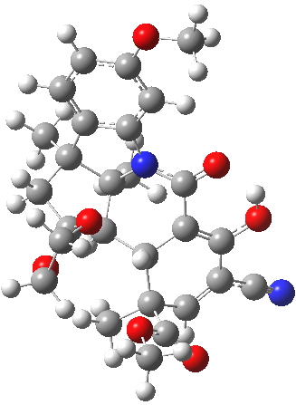
ΔG 0.0 kcal/mol ΔG +1.0 kcal/mol
H. Click for 3D

H. Click for 3D

J. Click for  3D.

J. Click for 3D.

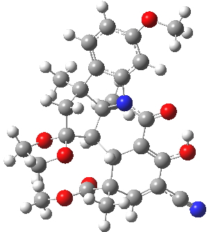
This computation shows that H is the lower in free energy by 1.0 kcal/mol, and by 0.8 kcal/mol in dispersion energy. So Woodward’s hypothesis that J was the more likely to form on steric grounds is not supported by this modern equivalent of a CPK model. I should add that a CPK model may only take an hour or so to build (but possibly weeks to order the components) whereas this quantum model took around 9 hours to compute. 

References

  1. E.J. Corey, and A.G. Hortmann, "The Total Synthesis of Dihydrocostunolide", Journal of the American Chemical Society, vol. 87, pp. 5736-5742, 1965. https://doi.org/10.1021/ja00952a037
  2. R.B. Woodward, and R. Hoffmann, "Stereochemistry of Electrocyclic Reactions", Journal of the American Chemical Society, vol. 87, pp. 395-397, 1965. https://doi.org/10.1021/ja01080a054
  3. H.S. Rzepa, "The Aromaticity of Pericyclic Reaction Transition States", Journal of Chemical Education, vol. 84, pp. 1535, 2007. https://doi.org/10.1021/ed084p1535

A comparison of left and right handed DNA double-helix models.

Saturday, January 1st, 2011

When Watson and Crick (WC) constructed their famous 3D model for DNA, they had to decide whether to make the double helix left or right handed. They chose a right-handed turn, on the grounds that their attempts at left-handed models all “violated permissible van der Waals contacts“. No details of what these might have been were given in their original full article (or the particular base-pairs which led to the observation). This follow-up to my earlier post explores this aspect, using a computer model.

One half of a (CGCG) DNA strand

The DNA model used here is shown above; in shorthand it is d(CGCG)2. A crystal structure reveals it to form a (non-Watson-Crick) left-handed helix. If you open the 3D model below (based on a ωB97XD/6-31G(d)/SCRF=water optimisation), some of the short van der Waals contacts are measured. Most are around 2.25Å and the shortest is 2.1Å. It is worth noting that WC note in their article that a distance of 2.1Å for the B-form is acceptable (p92, bottom) and not a violation. All twelve hydrogen bond lengths H…O or H…N are normal, with lengths around 1.8Å. Given that a H…H distance is at its most attractive at ~2.4Å, and plenty of H…H distances of ~2.1Å are known from the crystal structures of organic molecules, one might conclude that (for the CG base pair), their hypothesis that the Z-form could be eliminated was wrong.

The DNA duplex d(CGCG) showing a left handed helix with short H...H contacts shown. Click for 3D

But might the original WC-right handed form for this system be at least competitive? There is one H…H of 2.05Å and quite a few at ~2.5Å (3D model below). The “violation” of van der Waals contacts is if anything slightly worse than with the left-handed helix. The total difference in the dispersion energy is a rather astonishing ~12 kcal/mol in favour of the Z-form. I will update this post (as a comment) when the relative free energies of the two forms are available (this calculation takes a while), but there is little doubt that the Z-form is indeed the more stable.

The DNA duplex d(CGCG) showing a right handed helix with short H...H contacts shown. Click for 3D

What can also be said about the Watson-Crick right handed form is that the hydrogen bonding is not so optimal. One of the twelve interactions between a (terminal) CG pair has some signs of being “unzipped“, with an N-H…O=C distance of ~1.9Å (there is no sign of similar unzipping in the Z-form). One must wonder whether this difference in the Z- and B-helices for the CG pair has been exploited in nature.


One crucial aspect of DNA is the local conformation about the bond connecting the base and the ribose, N9-C8 in the diagram below(green arrow).

Conformation of the base-ribose unit

An analysis of this bond can be expressed in terms of NBO theory. This clearly shows a strong interaction energy (E2) between the lone pair on N9 and the C8-O4 antibonding orbital of 13.3 kcal/mol, a classical anomeric effectin fact. In this case, it promotes the local conformation of this unit, which has a significant effect on the final model.

What else can analysis of the wavefunction tell us? Well, curiously, the optical rotation of this particular small oligomer has never been reported in the literature, and an intriguing question is whether it might have proved useful to distinguish between B- and Z-forms of the duplex? To do this, one needs a reasonably reliable way of computing [α]D for both isomers. This is because optical rotations are not reliably additive, and it is difficult to estimate them accurately based purely on the fragments present in the molecule. In 2011, is is now perfectly possible to calculate this quantity quantum mechanically, even for 250 atoms, using a reasonable basis set and making allowance for solvation (which is known to affect the calculated rotation). The values (CAM-B3LYP/6-31G(d)/SCRF=water) for the Z-isomer are 66° and 32° for the B-isomer. Of course the model is not complete, lacking a counterion for the phosphate and explicit water molecules, but even so, it might appear that the reason optical rotations are not reported is that they truly are not useful!