Posts Tagged ‘Nature’

Dispersion-induced triplet aromatisation?

Thursday, January 3rd, 2019

There is emerging interest in cyclic conjugated molecules that happen to have triplet spin states and which might be expected to follow a 4n rule for aromaticity.[1] The simplest such system would be the triplet state of cyclobutadiene, for which a non or anti-aromatic singlet state is always found to be lower in energy. Here I explore some crystal structures containing this motif for possible insights.

My search query is shown below, and the search is constrained so that the four substituents are Si, C or H.


The results show three clusters. The top left and bottom right have one long bond length ~1.6Å and the other much shorter at ~1.35Å (Δr ~0.25Å) The central region contains two examples, 2 where the difference between the two lengths is rather smaller and 1 where they are equal.

The first example 1[2] is in fact the di-anion of cyclobutadiene and as a 6π aromatic, one indeed expects the C-C bonds to be equal in length. The second 2 is tetra t-butylcyclobutadiene as reported in 1983.[3] At room temperature the two C-C bond lengths are 1.464 and 1.483Å, at -30°C, 1.466 and 1.492Å and at -150°C 1.441 and 1.526Å (Δr 0.085Å). These results led to the conclusion that this species was not intrinsically square but rectangular, as expected of singlet cyclobutadiene. The equalisation was attributed to equal populations of two disordered rectangular orientations averaging to an approximately square shape at higher temperatures.

But why is the behaviour of this particular cyclobutadiene different from the others in the plot above? Perhaps the answer lies these in the results of the Schreiner group[4], in which the dispersion attractions of substituents such as t-butyl can have substantial and often unexpected effects on the structures of molecules. So it is reasonable to pose the question; could the room temperature bond length differences of 2 be smaller compared with the other more extreme examples as a result of dispersion effects?

Here I have computed the singlet geometry of tetra t-butylcyclobutadiene at the B3LYP+D3BJ/Def2-TZVPP level (i.e. using the D3BJ dispersion correction, FAIR data DOI: 10.14469/hpc/4924). Δr for this singlet state is 0.264Å, larger than apparently from the crystal structure, but in agreement with the other crystal results as seen above.

The origins of the measured structure of 2 must be in the barrier to the automerisation of the singlet state. For normal cyclobutadienes, this must be relatively high since the transition state is presumably anti-aromatic. High enough that the averaging of the two rectangular structures is slow enough that it manifests as two different bond lengths. But in 2, as the temperature of the crystal increases, the bonds become more equal, suggesting a lower barrier to the equalisation than the other examples. This is also supported by the apparent identification of a triplet square state for the tetra-TMS analogue of tetra-tert-butyl cyclobutadiene derivative [5] which again suggests that dispersion might favour a square form over the rectangular one.

To finish, I show the crystal structure search for the 8-ring homologue of cyclobutadiene, plotted for the two adjacent C-C lengths and (in colour) the dihedral angle associated with the three atoms involved and the fourth along the ring. Cluster 1 represents various boat-shaped derivatives with very different C-C bond lengths. Cluster 2 are all ionic, and as per above represent a planar 10π-electron ring. Cluster 3 are mostly “tethered” molecules in which additional rings enforce planarity. 

COT

Unfortunately, none of these derivatives include tert-butyl or TMS derivatives in adjacent positions around the central ring. Perhaps octa(t-Bu)cyclo-octatetraene or its TMS analogue would be interesting molecules to try to synthesize!

References

  1. A. Kostenko, B. Tumanskii, Y. Kobayashi, M. Nakamoto, A. Sekiguchi, and Y. Apeloig, "Spectroscopic Observation of the Triplet Diradical State of a Cyclobutadiene", Angewandte Chemie International Edition, vol. 56, pp. 10183-10187, 2017. https://doi.org/10.1002/anie.201705228
  2. T. Matsuo, T. Mizue, and A. Sekiguchi, "Synthesis and Molecular Structure of a Dilithium Salt of the <i>cis</i>-Diphenylcyclobutadiene Dianion", Chemistry Letters, vol. 29, pp. 896-897, 2000. https://doi.org/10.1246/cl.2000.896
  3. H. Irngartinger, and M. Nixdorf, "Bonding Electron Density Distribution in Tetra‐<i>tert</i>‐butylcyclobutadiene— A Molecule with an Obviously Non‐Square Four‐Membered ring", Angewandte Chemie International Edition in English, vol. 22, pp. 403-404, 1983. https://doi.org/10.1002/anie.198304031
  4. S. Rösel, H. Quanz, C. Logemann, J. Becker, E. Mossou, L. Cañadillas-Delgado, E. Caldeweyher, S. Grimme, and P.R. Schreiner, "London Dispersion Enables the Shortest Intermolecular Hydrocarbon H···H Contact", Journal of the American Chemical Society, vol. 139, pp. 7428-7431, 2017. https://doi.org/10.1021/jacs.7b01879

Open Access journal publishing debates – the elephant in the room?

Sunday, November 4th, 2018

For perhaps ten years now, the future of scientific publishing has been hotly debated. The traditional models are often thought to be badly broken, although convergence to a consensus of what a better model should be is not apparently close. But to my mind, much of this debate seems to miss one important point, how to publish data.

Thus, at one extreme is COAlition S, a model which promotes the key principle that “after 1 January 2020 scientific publications on the results from research funded by public grants provided by national and European research councils and funding bodies, must be published in compliant Open Access Journals or on compliant Open Access Platforms.” This includes ten principles, one of which “The ‘hybrid’ model of publishing is not compliant with the above principles” has revealed some strong dissent, as seen at forbetterscience.com/2018/09/11/response-to-plan-s-from-academic-researchers-unethical-too-risky I should explain that hybrid journals are those where the business model includes both institutional closed-access to the journal via a subscription charge paid by the library, coupled with the option for individual authors to purchase an Open Access release of an article so that it sits outside the subscription. The dissenters argue that non-OA and hybrid journals include many traditional ones, which especially in chemistry are regarded as those with the best impact factors and very much as the journals to publish in to maximise both the readership, hence the impact of the research and thus researcher’s career prospects. Thus many (not all) of the American Chemical Society (ACS) and Royal Society of Chemistry (RSC) journals currently fall into this category, as well as commercial publishers of journals such as Nature, Nature Chemistry,Science, Angew. Chemie, etc. 

So the debate is whether funded top ranking research in chemistry should in future always appear in non-hybrid OA journals (where the cost of publication is borne by article processing charges, or APCs) or in traditional subscription journals where the costs are borne by those institutions that can afford the subscription charges, but of course also limit the access.  A measure of how important and topical the debate is that there is even now a movie devoted to the topic which makes the point of how profitable commercial scientific publishing now is and hence how much resource is being diverted into these profit margins at the expense of funding basic science.

None of these debates however really takes a close look at the nature of the modern research paper. In chemistry at least, the evolution of such articles in the last 20 years (~ corresponding to the online era) has meant that whilst the size of the average article has remained static at around 10 “pages” (in quotes because of course the “page” is one of those legacy concepts related to print), another much newer component known as “Supporting information” or SI has ballooned to absurd sizes. It can reach 1000 pages[1] and there are rumours of even larger SIs. The content of SI is of course mostly data. The size is often because the data is present in visual form (think spectra). As visual information, it is not easily “inter-operable” or “accessible”. Nor is it “findable” until commercial abstracting agencies chose to index it. Searches of such indexed data are most certainly “closed” (again depending on institutional purchases of access) and not “open access”. You may recognise these attributes as those of FAIR (Findable, accessible, inter-operable and re-usable). So even if an article in chemistry is published in pure OA form, in order to get FAIR access to the data associated with the article, you will probably have to go to a non-OA resource run by a commercial organisation for profit. Thus a 10 page article might itself be OA, but the full potential of its 1000+ page data (an elephant if ever there was one) ends up being very much not OA.

You might argue that the 1000+ pages of data does not require the services of an abstracting agency to be useful. Surely a human can get all the information they want from inspecting a visual spectrum? Here I raise the future prospects of AI (artificial intelligence). The ~1000 page SI I noted above[1] includes e.g NMR spectra for around 70 compounds (I tried to count them all visually, but could not be certain I found them all). A machine, trained to identify spectra from associated metadata (a feature of FAIR), could extract vastly more information than a human could from FAIR raw data (a spectrum is already processed data, with implied information/data loss) in a given time. And for many articles, not just one. Thus FAIR data is very much targeted not only at humans but at the AI-trained machines of the future.

So I again repeat my assertion that focussing on whether an article is OA or not and whether publishing in hybrid journals is to be allowed or not by funders is missing that 100-fold bigger elephant in the room. For me, a publishing model that is fit for the future should include as a top priority a declaration of whether the data associated with it is FAIR. Thus in the Plan-S ten principles, FAIR is not mentioned at all. Only when FAIR-enabled data becomes part of the debates can we truly say that the article and its data are on its way to being properly open access.


The FAIR concept did not originally differentiate between processed data (i.e. spectra) and the underlying primary or raw data on which the processed data is based. Our own implementation of FAIR data includes both types of data; raw for machine reprocessing if required, and processed data for human interpretation. Along with a rich set of metadata, itself often created using carefully designed workflows conducted by machines.

The proportion of articles relating to chemistry which do not include some form of SI is probably low. These would include articles which simply provide a new model or interpretation of previously published data, reporting no new data of their own. A famous historical example is Michael Dewar’s re-interpretation of the structure of stipitatic acid[2] which founded the new area of non-benzenoid aromaticity.

References

  1. J.M. Lopchuk, K. Fjelbye, Y. Kawamata, L.R. Malins, C. Pan, R. Gianatassio, J. Wang, L. Prieto, J. Bradow, T.A. Brandt, M.R. Collins, J. Elleraas, J. Ewanicki, W. Farrell, O.O. Fadeyi, G.M. Gallego, J.J. Mousseau, R. Oliver, N.W. Sach, J.K. Smith, J.E. Spangler, H. Zhu, J. Zhu, and P.S. Baran, "Strain-Release Heteroatom Functionalization: Development, Scope, and Stereospecificity", Journal of the American Chemical Society, vol. 139, pp. 3209-3226, 2017. https://doi.org/10.1021/jacs.6b13229
  2. M.J.S. DEWAR, "Structure of Stipitatic Acid", Nature, vol. 155, pp. 50-51, 1945. https://doi.org/10.1038/155050b0

Organocatalytic cyclopropanation of an enal: (computational) assignment of absolute configurations.

Saturday, September 1st, 2018

I am exploring the fascinating diverse facets of a recently published laboratory experiment for undergraduate students.[1] Previously I looked at a possible mechanistic route for the reaction between an enal (a conjugated aldehyde-alkene) and benzyl chloride catalysed by base and a chiral amine, followed by the use of NMR coupling constants to assign relative stereochemistries. Here I take a look at some chiroptical techniques which can be used to assign absolute stereochemistries (configurations).

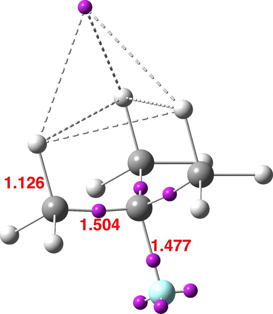
I will focus on the compound 4a, the major stereochemical product of this student laboratory reaction, with the stereochemistry as represented in e.g. the abstract of the main article[1] and shown below with added CIP (Cahn-Ingold-Prelog) notation as (1S,2R,3R);

Its enantiomer (not shown in the article) is of course;

In the article supporting information[1]), the major diasteromer of 4a deriving from use of the S stereoisomer of the prolinol catalyst is reported as having an optical rotation (ORP) [α]D25 of -62.4°, p6 or -58.1°, p5), but the stereo-labels are not added there. On  p1 (“based on a student report”) 4a was however labelled as (1R,2S,3S) and the chirality (S) of the catalyst used was also noted in the adjacent experimental procedure. One might then reasonably match (1R,2S,3S)-4a to the S-catalyst and hence (1S,2R,3R)-4a to the R-catalyst.  However, in a laboratory environment where both S and R catalysts are in circulation, it is always useful to have procedures available for independent checks.

There are two methods of assigning absolute chirality, crystallography and chiroptical spectroscopy. The former does require crystalline samples; the latter can use solutions. To cut to the chase, the former method was used for a related compound where the n-heptyl group above is replaced by a p-chlorophenyl substituent (perhaps because the latter imparts suitable crystallinity). On p S123 of the SI of an earlier article[2] the assignment for the p-chlorophenyl derivative was as (1R,2S,3S)-4a for S-catalyst (see DOI: 10.5517/ccdc.csd.cc1mcqg5 OZAXEU). But this procedure is not entirely foolproof; the stereochemistry is decided by interactions between often bulky substituents at the transition state and it might be that here the p-chlorophenyl derivative has different properties from n-heptyl. Moreover bulk solutions may be different in their composition from single crystals. So it is useful to obtain independent proof.

An absolute assignment procedure based on chiroptical methods was first famously used by Kirkwood in 1951 (the Fischer convention is confirmed as a structurally correct representation of absolute configuration).[3] Such calculations need to take into account e.g. rotational conformers about the two bonds labelled in red above. In the previous post, I had noted variation of up to 2Hz in the calculated 3JHH coupling constants as a result of this mobility. This variation is probably too small to really influence any relative stereochemical interpretations, but is the same true for chiroptical assignments?

In Table 1 we can see whether this is still true for the predicted optical rotation of compound 4a, using two different functionals for the calculation (B3LYP and M062X respectively). The results rather surprised me; a simple bond rotation of an aryl or carbonyl group can invert the sign of the rotation. Clearly the observed optical rotation of -62.4° arises from a suitable combination of different Boltzmann populations of the individual bond rotamers, but to combine these accurately you would need to know the solution populations themselves very accurately and that is quite a challenge. So at this stage, we do not really have totally convincing independent evidence of whether the observed negative optical rotation corresponds to (1S,2R,3R)-4a or to its enantiomer (1R,2S,3S).

Table 1. Calculated Optical rotations for (1S,2R,3R)-4a. 

FAIR Data DOI: 10.14469/hpc/4678

Conformer

ORP [α]D, B3LYP+GD3BJ/Def2-TZVPP/SCRF=chloroform

ORP [α]D, M062X/Def2-TZVPP/SCRF=chloroform

4 +376 +238
3 -335 -301
2 -247 -223
1 +710 +522

Next, another chiroptical technique, electronic circular dichroism, or ECD. Here, the sign of the difference in absorption of polarized light (Δε), and known at the Cotton effect, characterises the specific enantiomer. The experimental Cotton effect for compound 4a obtained from S-catalyst (known as 3d in the SI, p S142[2]) can be simply summarised as +ve@315nm and -ve@275nm. Comparison with calculated spectra (Figure S17, p S146-7[2])  was performed using a Boltzmann-averaging (albeit based on enthalpies rather than the formally correct free energies), for three significant populations and this procedure matched to (1R,2S,3S).  Since the reported calculations were apparently for gas phase (and replacing n-heptyl with methyl) here I have repeated them in the actual solvent used (acetonitrile) and with the heptyl present. Although the ECD responses can still be severely dependent on the conformation, three of the spectra qualitatively agree that the responses at ~300nm and 260 nm are respectively -ve and +ve. This confirms that (1S,2R,3R)-4a is the wrong enantiomer for S-catalyst and that the correct assignment is therefore (1R,2S,3S), as was indeed reported.[2]

Table 2. Calculated electronic circular dichroism for

 (1S,2R,3R)-4a. FAIR Data DOI: 10.14469/hpc/4678

Conformer

ECD calculation, ωB97XD/Def2-TZVPP

4
3
2
1

It is still true that the overall the fit between chiroptical experiment and theory can be sensitive to the Boltzmann population, as obtained from e.g. ΔΔG = -RT ln [1]/[2]), where 1 and 2 are two different conformers. ΔΔG is a difficult energy difference to compute accurately. Here is a suggested exercise in the statistics of error propagation. How does an error in ΔΔG propagate to the ratio of concentrations of two conformers [1]/[2]? Or, how accurately must ΔΔG be calculated in order to predict conformer populations to say better than 5%.

One more go at chiroptics, this time Vibrational Circular Dichroism, or VCD. The nature of the chromophore is different, but the principle is the same as ECD. I have deliberately truncated the spectrum to cut off all vibrations below 1000 cm-1 (these being the modes associated with group rotations) but to no avail, the four conformations all still look too different to avoid doing a Boltzmann averaging.

Table 3. Calculated VCD spectra for (1S,2R,3R)-4a. 

Conformer Spectrum
4
3
2
1

A modern VCD instrument does have one trick up its sleeve for coping with the conformer problem. The sample (as a thin-film) can be annealed down to very low temperatures before the spectrum is recorded. This effectively removes all higher energy forms, leaving just the most stable conformation as the only species present. However, that is an expensive experiment (and instrument!) to use.

There are perhaps some 2 million scalemic molecules (substances where one chiral form is in excess over the mirror image) for which chiroptical properties have been reported, but probably <50,000 crystal structures where absolute configurations have been assigned. Thus the vast majority of absolute configuration assignments have been done either chiroptically or by synthetic correlations (chemical transformations from molecules of known absolute configuration, with the assumption that you know how each transformation affects the chiral centres present). Given some of the difficulties and challenges noted above, it is tempting to conclude that a significant proportion of those 2 million molecules may have been mis-assigned (I once estimated up to 20%). However, we may conclude that the molecules discussed here are safely assigned correctly! 


No CIP-stereolabels appear in the article itself.[1] Perhaps this assignment is omitted in order to provide a student exercise? There are many errors in stereochemical assignments in the literature. A good many of them may be the result of simple sample mis-labelling.[4] The caption to Figure S17 states All the simulations are for the 1R,2R,3S absolute configuration. This is probably an error and should read 1R,2S,3SA correction of ~+15nm is sometimes applied to these values, but not done here.


References

  1. M. Meazza, A. Kowalczuk, S. Watkins, S. Holland, T.A. Logothetis, and R. Rios, "Organocatalytic Cyclopropanation of (<i>E</i>)-Dec-2-enal: Synthesis, Spectral Analysis and Mechanistic Understanding", Journal of Chemical Education, vol. 95, pp. 1832-1839, 2018. https://doi.org/10.1021/acs.jchemed.7b00566
  2. M. Meazza, M. Ashe, H.Y. Shin, H.S. Yang, A. Mazzanti, J.W. Yang, and R. Rios, "Enantioselective Organocatalytic Cyclopropanation of Enals Using Benzyl Chlorides", The Journal of Organic Chemistry, vol. 81, pp. 3488-3500, 2016. https://doi.org/10.1021/acs.joc.5b02801
  3. W.W. Wood, W. Fickett, and J.G. Kirkwood, "The Absolute Configuration of Optically Active Molecules", The Journal of Chemical Physics, vol. 20, pp. 561-568, 1952. https://doi.org/10.1063/1.1700491
  4. H.S. Rzepa, "The Chiro-optical Properties of a Lemniscular Octaphyrin", Organic Letters, vol. 11, 2009. https://doi.org/10.1021/ol901172g

Why do flowers such as roses, peonies, dahlias, delphiniums (etc), exhibit so many shades of colours?

Monday, June 18th, 2018

It was about a year ago that I came across a profusion of colour in my local Park. Although colour in fact was the topic that sparked my interest in chemistry many years ago (the fantastic reds produced by diazocoupling reactions), I had never really tracked down the origin of colours in many flowers. It is of course a vast field. Here I take a look at just one class of molecule responsible for many flower colours, anthocyanidin, this being the sugar-free counterpart of the anthocyanins found in nature.

These vary widely in the substituent around the aromatic rings, but here I take a look at just three differing substitutions. Thus pelargonidin has just one OH on ring C (R1‘, R3‘=H, see crystal structure[1]), cyanidin has two (R5‘=H, see crystal structure[2]) and is found in red roses, dahlia, peonies etc. Finally delphinidin (no crystal structure available) has three OHs in that region and is found in yes, delphiniums but also grape skins etc. Below is a colour table that allows one to relate the electronic transitions in a molecule to the observed colour, which of course is due to removal (absorption) of wavelength of light leaving us to see all the remaining wavelengths.
colour table

Next I show the computed UV/visible spectra of these three species (ωB97XD/6-311G(d,p)/SCRF=water). Click on any image to se a 3D model of the molecule.

Note how in the visible region, all have a very simple (monochromatic) single electronic transition comprising mostly the HOMO→LUMO excitation.

Click to view 3D model of the HOMO

Click to view 3D model of the LUMO

Now, λmax can be predicted quite poorly using most DFT methods, but the trends should be better predicted. Thus the change induced by adding two hydroxy groups is ~7nm, which is in effect how the colour seen in a flower can be tuned to display different shades.

Next, pH. Using delphinidin, under basic conditions one can remove a proton from the cationic species to produce a neutral quinone. In fact, any one of five OH groups could have its proton removed and so it is of some interest to compare the relative energies of the five isomers so produced.

Position proton removed Relative ΔG298, kcal/mol
4′ 0.0
5 3.8
7 4.7
3′ 11.8
5′ 22.2

In fact, one species only would have the major Boltzmann population (4′) and so we need only look at its UV/Visible predicted spectrum. This is shifted 17nm towards the red, thus producing a blue colour in what remains after it is absorbed. The absorption (ε) also increases significantly. Indeed the very striking colour of blue delphiniums (one of my favourite flowers) must be produced by such pH control in the plant. Given the presence of delphinidin in many grape skins, the next time I drink a glass of red wine, I will see if it turns blue upon adding some NaOH!


FAIR data doi: 10.14469/hpc/4473

References

  1. N. Saito, and K. Ueno, "The Crystal and Molecular Structure of Pelargonindin Bromide Monohydrate", HETEROCYCLES, vol. 23, pp. 2709, 1985. https://doi.org/10.3987/r-1985-10-2709
  2. K. Ueno, and N. Saito, "Cyanidin bromide monohydrate (3,5,7,3',4'-pentahydroxyflavylium bromide monohydrate)", Acta Crystallographica Section B Structural Crystallography and Crystal Chemistry, vol. 33, pp. 114-116, 1977. https://doi.org/10.1107/s0567740877002702

A record polarity for a neutral compound?

Friday, April 13th, 2018

In several posts a year or so ago I considered various suggestions for the most polar neutral molecules, as measured by the dipole moment. A record had been claimed[1] for a synthesized molecule of ~14.1±0.7D. I pushed this to a calculated 21.7D for an admittedly hypothetical and unsynthesized molecule. Here I propose a new family of compounds which have the potential to extend the dipole moment for a formally neutral molecule up still further.

These molecules derive from a well-known class of molecule known as ortho-quinomethides. If the methide part is substituted with an electron donating substituent such as an amino group in 3, a push-pull opportunity now arises, which is strongly driven by aromatisation of the quinomethide ring. This allows one to design “neutral” molecules such as 1 and 2, which now contain respectively two and three rings that will be aromatised by the process. The aromatisation stabilization energy is balanced of course by an opposing increase in energy resulting from charge separation. You can observe that partially aromatising three independent rings as in 2 can drive a great deal of charge separation. One may indeed wonder how much charge separation can be sustained before a triplet instability occurs, driving the molecule back to being neutral. In the case of 2, the wavefunction is in fact stable to such an open shell state, but higher homologues may not be. An aspect worth investing!

1 2
DM 12.3 DM 31.5
DOI: 10.14469/hpc/4004 DOI: 10.14469/hpc/4059

Molecule 1 does have some precedent in 3[2] but this system exists as a phenol, having abstracted a proton from an acid and leaving behind the acid anion, as per below for 1. Any attempts to deprotonate this phenol with a superstrong base were unreported.

Unsurprisingly therefore, molecules such as 1 and 2 could be regarded as even more highly potent bases than 3, driven again by further aromatisation. The properties of such a potential superbase will be investigated in the next post.

References

  1. J. Wudarczyk, G. Papamokos, V. Margaritis, D. Schollmeyer, F. Hinkel, M. Baumgarten, G. Floudas, and K. Müllen, "Hexasubstituted Benzenes with Ultrastrong Dipole Moments", Angewandte Chemie International Edition, vol. 55, pp. 3220-3223, 2016. https://doi.org/10.1002/anie.201508249
  2. N.R. Candeias, L.F. Veiros, C.A.M. Afonso, and P.M.P. Gois, "Water: A Suitable Medium for the Petasis Borono‐Mannich Reaction", European Journal of Organic Chemistry, vol. 2009, pp. 1859-1863, 2009. https://doi.org/10.1002/ejoc.200900056

What are the highest bond indices for main group and transition group elements?

Sunday, March 4th, 2018

A bond index (BI) approximately measures the totals of the bond orders at any given atom in a molecule. Here I ponder what the maximum values might be for elements with filled valence shells.

Following Lewis in 1916[1] who proposed that the full valence shell for main group elements should be 2 (for the first two elements) and 8 (the “octet“), Bohr (1922[2]), Langmuir (1919-1921[3]) and Bury (1921[4]) extended this rule to include 18 (the transition series) and 32 (the lanthanides and actinides). If we assume no contributions from higher Rydberg shells (thus 3s, 3p, 3d for carbon etc) and an electron pair model for orbital population (which amounts to the single-determinantal model), then the maximum bond index for hydrogen (and helium) would be 1, it would be 4 for main group elements, and then what?

For the special case of hydrogen, I have previously identified (for a hypothetical species) a bond index of 1.33, due mostly to a high Rydberg occupancy of 1.19e. The more normal BI is <1.0, as noted for this hexacoordinated hydride system. My current estimate for the maximum bond index for main group elements is <4.5. Thus for SF6, it has the value of ~4.33 and that includes a modest occupancy of Rydberg shells of 0.36e = 0.18 BI. Exclude these and it is close to 4.

Move on from group 16 to group 6 and you get compounds such as Me4CrCrMe44- or ReMe82- where the metal bond indices are ~6.5. Compounds such as Cr(Me)6 (BI = 5.6)  and W(Me)(BI = 6.1) are rather lower. This is a long way from 18/2 = 9. The lanthanides and actinides[5] are unlikely to reveal many large BIs (32/2= 16 maximum value) since they are often ionic and the wavefunctions may be too complex to allow a simple index such as a BI to be safely computed.

So if we are hunting for record BIs, the transition elements are the place to hunt. Can a BI of 6.5 be beaten? Can it even approach 9, its maximum value? Does anyone know of candidate molecules? 


FAIR Data doi: 10.14469/hpc/3352.

References

  1. G.N. Lewis, "THE ATOM AND THE MOLECULE.", Journal of the American Chemical Society, vol. 38, pp. 762-785, 1916. https://doi.org/10.1021/ja02261a002
  2. N. Bohr, "Der Bau der Atome und die physikalischen und chemischen Eigenschaften der Elemente", Zeitschrift f�r Physik, vol. 9, pp. 1-67, 1922. https://doi.org/10.1007/bf01326955
  3. I. Langmuir, "Types of Valence", Science, vol. 54, pp. 59-67, 1921. https://doi.org/10.1126/science.54.1386.59
  4. C.R. Bury, "LANGMUIR'S THEORY OF THE ARRANGEMENT OF ELECTRONS IN ATOMS AND MOLECULES.", Journal of the American Chemical Society, vol. 43, pp. 1602-1609, 1921. https://doi.org/10.1021/ja01440a023
  5. P. Pyykkö, C. Clavaguéra, and J. Dognon, "The 32‐Electron Principle", Computational Methods in Lanthanide and Actinide Chemistry, pp. 401-424, 2015. https://doi.org/10.1002/9781118688304.ch15

Hypervalent or not? A fluxional triselenide.

Saturday, February 24th, 2018

Another post inspired by a comment on an earlier one; I had been discussing compounds of the type I.In (n=4,6) as possible candidates for hypervalency. The comment suggests the below as a similar analogue, deriving from observations made in 1989.[1]

This compound was investigated using 77Se NMR, with the following conclusions:

  1. The compound is fluxional, with the lines at room temperature broadened compared to those at -50°C.
  2. At -50°C the peaks are sharp enough to discern 1JSe-Se couplings, with multiplicities and integrations that suggest a central Se is surrounded by four equivalent further Se atoms, with shifts of 655.1 and 251.2 ppm.
  3. The magnitude of this 1JSe-Se coupling (391 Hz) leads to the suggestion of a considerable contribution of a resonance form with Se=Se bonds (structure 2 above).
  4. This was supported by 2J13C-77Se couplings which also imply a symmetrically coordinated central  Se.
  5. Thus the two resonance forms 1 or 2 above were suggested as the predominant form at -50°C, with an increasing incursion of the open chain isomer 3 at higher temperatures giving rise to the observed fluxional dynamic behaviour.
  6. One may surmise from these results that the central Se is certainly hypercoordinated and by the classical interpretations hypervalent.

Here are some calculations (R=H), at the ωB97XD/Def2-TZVPP/SCRF=chloroform level.‡ In red are the calculated Wiberg Se-Se bond orders, which give little indication of any Se=Se double bond character. 

The calculated 77Se shifts are shown in magenta, with the observed values being 655 and 255 ppm. The match is not good, the errors were 120 and 20.5 ppm.  However calculated shifts for elements adjacent to e.g. Se or Br etc suffer from relativistic effects such as spin orbit coupling.[2] Thus the shift for the central Se, surrounded by four other Se atoms is likely to have a significant error, but the error for the four other Se atoms should be less. The reverse is true.

However, all the calculations of this species (up to Def2-TZVPPD basis set) showed this symmetric form of D2h symmetry to actually be a transition state, as per below.

There is a minimum with the structure below in which one pair of Se-Se lengths are longer than the other pair and for which the free energy is 6.5 kcal/mol lower. The Wiberg bond orders for the two sets of Se-Se bonds are now 0.16 and 0.86, which very much corresponds to structure 3 above.

Assuming that this compound is fluxional even at -50°C, the average of the pairs of Se atoms gives calculated shifts of 667 ppm (655 obs) whilst the central Se is 204.6 ppm (251 obs). The latter, influenced by two especially short Se-Se distances, is likely to have a very large spin-orbit coupling error, whilst for the former the error will be smaller (13C shifts adjacent to one Br typically have induced calculated errors of about 14 ppm[2]).

At this point I searched the Cambridge structure database for Se coordinated by four other Se atoms. A close analogue[3] has the structure shown below, in which pairs of Se-Se interactions have unequal bond lengths, the shorter being ~2.45Å. This matches the calculation above reasonably well.

Reconciling these various observations, we might assume that even at -50°C the fluxional behaviour has not been frozen out. Given that the fluxional barrier is only 6.5 kcal/mol, it is unlikely that the spectrum could be measured at a sufficiently low temperature to reveal not two sets of Se signals in the ratio 4:1 but three in the ratio 2:2:1. The spin-spin couplings reported presumably are a result of averaging a genuine 1JSe-Se coupling with a through space coupling.

So it appears that the analysis of the 77Se NMR reported in this article [1] may not be quite what it seems. A better interpretation is that structure 3 is the most realistic. This means no hypercoordination for the Se, never mind hypervalence!


FAIR data at DOI: 10.14469/hpc/3724. The original reference, Me2Se was incorrectly calculated without solvation by chloroform. The values shown here are now corrected from those shown in the original post.

References

  1. Y. Mazaki, and K. Kobayashi, "Structure and intramolecular dynamics of bis(diisobutylselenocarbamoyl) triselenide as identified in solution by the 77Se-NMR spectroscopy", Tetrahedron Letters, vol. 30, pp. 2813-2816, 1989. https://doi.org/10.1016/s0040-4039(00)99132-9
  2. D.C. Braddock, and H.S. Rzepa, "Structural Reassignment of Obtusallenes V, VI, and VII by GIAO-Based Density Functional Prediction", Journal of Natural Products, vol. 71, pp. 728-730, 2008. https://doi.org/10.1021/np0705918
  3. R.O. Gould, C.L. Jones, W.J. Savage, and T.A. Stephenson, "Crystal and molecular structure of bis(NN-diethyldiselenocarbamato)-selenium(II)", Journal of the Chemical Society, Dalton Transactions, pp. 908, 1976. https://doi.org/10.1039/dt9760000908

Hypervalent hydrogen?

Saturday, January 13th, 2018

I discussed the molecule the molecule CH3F2- a while back. It was a very rare computed example of a system where the added two electrons populate the higher valence shells known as Rydberg orbitals as an alternative to populating the C-F antibonding σ-orbital to produce CH3 and F. The net result was the creation of a weak C-F “hyperbond”, in which the C-F region has an inner conventional bond, with an outer “sheath” encircling the first bond. But this system very easily dissociates to CH3 and F and is hardly a viable candidate for experimental detection.  In an effort to “tune” this effect to see if a better candidate for such detection might be found, I tried CMe3F2-. Here is its story.

The calculation is at the ωB97XD/Def2-TZVPPD/SCRF=water level (water is here used as an approximate model for a condensed environment, helping to bind the two added electrons).

  1. An NBO (Natural Bond orbital) analysis reveals a total Rydberg orbital population of 1.186e and the following bond indices; F 0.853, C 3.977, C(methyl) 4.051, H(*3) 1.332. The latter corresponds to the three methyl hydrogens aligned antiperiplanar to the C-F bond.
  2. To put this value into context, the hydrogen in the FHF anion has an NBO H bond index of 0.724, and the bridging hydrogens in diborane only have a value of 0.988. Even the hexa-coordinate hydride system [Co6H(CO)15] discussed in an earlier blog  has an H bond index of just 0.86. Actually, coordination of six or even higher for hydrogen is no longer rare; some 28 crystal structures of the type HM6 (M=metal) are known (it would be useful to find out if any of the other 27 such structures might have a hydrogen bond index >1).
  3. Next, the ELF analysis (Electron localisation function), analysed firstly using the excellent MultiWFN program.[1]

    This reveals an attractor basin integrating to 1.663e and located along the axis of the F-C bond and extended into the region of the three antiperiplanar methyl hydrogens. The C-F bond itself only supports a basin of 0.729e, typical of the fairly ionic C-F bond. The covalent C-Me bonds are also pretty normal, as are the other hydrogens.
  4. I also show ELF analysis using the alternative TopMod program[2]; the numerical values on this diagram are the calculated bond lengths in Å. The basin integrations are very similar to those obtained using MultiWFN.

    The Wiberg bond orders of the three H…H regions shown connected by dashed lines above are 0.154, which contributes to the bond index of >1 at these three hydrogens.
  5. The predicted 1H chemical shift of these three “hypervalent” hydrogens is +3.0 ppm, whilst the other six methyl hydrogens are at -0.87ppm.

So changing CH3F2- to CMe3F2- has dramatically changed the bonding picture that emerges, rather than a fine-tuning. The C-F is no longer a “hyperbond”, although the Rydberg occupancy of 1.186e remains unusually large. Most of the additional electrons have fled the torus surrounding the C-F bond and relocated to the exo-region of that bond where they now influence the three antiperiplanar methyl hydrogens. A two-electron-three-centre interaction if you like, but with the electron basin occupying a tetrahedral vertex rather than the triatom centroid.

I end with a challenge. Is it possible to find “real” molecules containing hydrogen where the formal bond index for at least one hydrogen exceeds 1.0 significantly, thus making it hypervalent? 


The calculations are all collected at FAIR doi; 10.14469/hpc/3372.

References

  1. T. Lu, and F. Chen, "Multiwfn: A multifunctional wavefunction analyzer", Journal of Computational Chemistry, vol. 33, pp. 580-592, 2011. https://doi.org/10.1002/jcc.22885
  2. S. Noury, X. Krokidis, F. Fuster, and B. Silvi, "Computational tools for the electron localization function topological analysis", Computers & Chemistry, vol. 23, pp. 597-604, 1999. https://doi.org/10.1016/s0097-8485(99)00039-x

Multispectral Chiral Imaging with a Metalens.

Saturday, January 6th, 2018

The title here is from an article on metalenses[1] which caught my eye.

Metalenses are planar and optically thin layers which can be manufactured using a single-step lithographic process. This contrasts with traditional lenses that are not flat and where the optical properties result from very accurately engineered curvatures, which in turn are expensive to manufacture. Metalenses can have built into them many interesting optical properties, including light polarisation and dispersion. Nanoengineering has now resulted[1] in a metalens which can simultaneously present two images of opposite helicity of an object within the same field of view.

What is the relevance to chemistry? Well, a well-known chiroptical technique is known as electronic circular dichroism (ECD). At its simplest, it probes the difference in absorption by a chiral molecule of UV and visible light with opposite circular polarisation. This difference plotted as a function of the wavelength of the light is known as the ECD response. Importantly, this response can also be calculated for either enantiomer of the chiral molecule and hence the absolute configuration can be assigned on the basis of which calculated response matches the observed spectrum. Because the difference in response to the two polarisations of the light (Δε) is actually very small, the ECD technique is intrinsically less sensitive than e.g. normal UV/Visible spectra and this requires the use of expensive instruments to record that small difference. Chiral metalenses offer an interesting future opportunity to create new forms of ECD instrument, perhaps ones that are far more sensitive. In turn, this could lower the costs of acquiring ECD functionality in the standard laboratory (see [2] for an application in teaching laboratories). Very possibly, the most expensive component would in fact then be the computational simulations required to match up with the experimental spectrum!

When metalenses were first introduced, they were only able to lens a limited range of wavelengths. In another article by the same group[3] they now announce a new generation of metalens that covers the region 470 to 670 nm. This excludes the UV regions (<300nm) or the IR regions (>1200nm). The latter covers another important chiroptical instrumental technique known as vibrational circular dichroism, or VCD. As with ECD, the VCD response of a chiral molecule can be pretty well calculated using quantum chemistry and indeed often the VCD method is the only one that can successfully be used to assign absolute molecular configurations.[4] Unfortunately, VCD instruments are even more expensive than ECD ones, again largely due to the intrinsic insensitivity and the need to accumulate data using Fourier Transform methods over many hours. Few chemistry departments have such an instrument. So I will keep an eye out for when an effective chiral metalens operating in infra-red regions is announced! The prospect of routine VCD analyses is tantalising! 


References

  1. M. Khorasaninejad, W.T. Chen, A.Y. Zhu, J. Oh, R.C. Devlin, D. Rousso, and F. Capasso, "Multispectral Chiral Imaging with a Metalens", Nano Letters, vol. 16, pp. 4595-4600, 2016. https://doi.org/10.1021/acs.nanolett.6b01897
  2. K.K.(. Hii, H.S. Rzepa, and E.H. Smith, "Asymmetric Epoxidation: A Twinned Laboratory and Molecular Modeling Experiment for Upper-Level Organic Chemistry Students", Journal of Chemical Education, vol. 92, pp. 1385-1389, 2015. https://doi.org/10.1021/ed500398e
  3. W.T. Chen, A.Y. Zhu, V. Sanjeev, M. Khorasaninejad, Z. Shi, E. Lee, and F. Capasso, "A broadband achromatic metalens for focusing and imaging in the visible", Nature Nanotechnology, vol. 13, pp. 220-226, 2018. https://doi.org/10.1038/s41565-017-0034-6
  4. J.I. Murray, N.J. Flodén, A. Bauer, N.D. Fessner, D.L. Dunklemann, O. Bob‐Egbe, H.S. Rzepa, T. Bürgi, J. Richardson, and A.C. Spivey, "Kinetic Resolution of 2‐Substituted Indolines by <i>N</i>‐Sulfonylation using an Atropisomeric 4‐DMAP‐<i>N</i>‐oxide Organocatalyst", Angewandte Chemie International Edition, vol. 56, pp. 5760-5764, 2017. https://doi.org/10.1002/anie.201700977

Hypervalence and octet-expansion in trimethylene-λ6-sulfane and related species.

Monday, November 27th, 2017

Previously: “Non-polar” species such as SeMe6, SMe6, ClMe3, ClMe5 all revealed interesting properties for the Se-C, S-C or Cl-C “single” bonds. The latter two examples in particular hinted at internal structures for these single bonds, as manifested by two ELF basins for some of the bonds. Here I take a look at the related molecule where a formal double bond between carbon and the central sulfur atom replacing the single-bond might also hint at octet expansions and hypervalence.

Starting with X=Y=Z=CH2, the calculated (ωB97Xd/Def2-TZVPP) geometry has an interesting chiral D3-symmetric form. The density based ELF-basin centroids are shown below, with each formal C=S π-double bond represented by two ELF basins above and below the C-S axis and with each pair of ELF basins being twisted by 48° with respect to the other two pairs. The total valence shell count around the S is 10.98e and the octet is “expanded” (by ~3e).

The orbital-based NBO approach indicates little utilisation of higher (Rydberg) atomic orbital shells (S: [core]3S(1.13)3p(3.35)3d(0.11)4p(0.02); C: [core]2S(1.15)2p(3.77)3p(0.01)3d(0.01) ). Each S-C bond has a Wiberg bond order of 1.36 (significantly less than a double bond), and the central S has an overall bond index of 4.102. There is a real mis-match between the orbital partitioning (2*1.36 = 2.72e) and the ELF partitioning (2*1.83 = 3.66e) into the S-C bonds. The former indicates that ~two of the twelve valence electrons are entering into anti-bonding orbitals to reduce the total bond index from a possible six to just four, but that they still contribute to the electron-density based ELF disynaptic C-S basins. To cast light on this behaviour, successively one to three of the CH2 groups can be replaced by O.

For each “S=O” bond, we find the ELF basin population more or less halves and electrons instead populate the non-bonding O “lone pairs”. The S-C ELF populations in contrast remain approximately constant. These species therefore have “double” S=C bonds but just “single” S-O bonds. The Rydberg population increases slightly; S: [core]3S(1.06)3p(2.95)3d(0.16)4p(0.02)) and the S bond index is 4.18 for one oxygen and S: [core]3S(0.99)3p(2.67)3d(0.19)4p(0.02) and S bond index 4.16 for two oxygens.

Sulfur trioxide (below) seems best represented by S-O rather than S=O bonds. The Rydberg population is S: [core]3S(0.91)3p(2.41)3d(0.21)4p(0.03) and the S bond index is 4.32.

Just for good measure sulfur trisulfide S(S)3 shows rather lower lone pair population because of course it is less electronegative than oxygen, and hence has a slightly greater S-S ELF basin population. Rydberg, S: [core]3S(1.43)3p(3.73)3d(0.21)4p(0.03) and central S bond index 4.04.

It seems molecules where the electrons in a valence shell exceed the “octet” are only too happy to let the excess electrons leak out into adjacent electronegative atoms as lone pairs, where they are no longer classified as  “shared”. Trimethylene-λ6-sulfane does not have this option and the excess electrons remain in the region of the valence shell, but here they do not contribute to augmenting the bond index at the central atom.  In this specific interpretation, the octet is exceeded, but hypervalence is not induced. It is a slippery concept; one where general agreement about its properties may indeed be difficult to achieve!


The FAIR data DOI collection for this post is 10.14469/hpc/3316.