Posts Tagged ‘energy’
Wednesday, May 25th, 2011
This molecule is not leaving me in peace. It and I first met in 1990 (DO: 10.1039/C39910000765), when we spotted the two unusual π-facial bonds formed when it forms a loose dimer. The next step was to use QTAIM to formalise this interaction, and this led to spotting a second one missed the first time round (labelled 2 in that post). Then a method known as NCI was tried, which revealed an H…H interaction, labelled ? in that post! Here I discuss the origins of the ?

The Pirkle reagent
What sparked this re-visitation? Firstly, in this post, a CH…O interaction in Z-DNA was identified using NCI, and its origins probed using NBO E2 perturbation energies, which revealed that the C-H bond was antiperiplanar to a C-O bond (effects 2 and 3 in that post), that could have the effect of acidifying the H, and making it more prone to hydrogen bond to the lone pair of an oxygen. Inspired by this, I worked out how to display NCI colour codes surfaces here on this blog using Jmol (previously, VMD had been used, which cannot be embedded in a blog).

NCI Surface for the Pirkle reagent. Click for 3D
The H..H interaction previously alluded to is shown with a magenta arrow (between the two atoms with halos in the 3D model). The colour coding
blue means it is distinctly attractive. In my (second) post, I did not mention why it might be special (and also the colour coding covered a large range, which meant the blue tinge did not really stand out visually). Why might that interaction be significant? Well, the C-H bond is perfectly aligned with the C-F σ* orbital. The NBO E2 energy is 3.9 kcal/mol, which represents a modest acidification of the hydrogen.
You may notice from the above other blue regions. Click on the diagram, and go explore them. The history of this molecule is such that it is bound to hold more surprises!
Tags:energy, Julia Contreras-Garcia, Tutorial material
Posted in Interesting chemistry | 1 Comment »
Wednesday, May 18th, 2011
In earlier posts, I alluded to what might make DNA wind into a left or a right-handed helix. Here I switch the magnification of our structural microscope up a notch to take a look at some more inner secrets.

A fragment of a single chain of DNA, taken from a Z-helix. Click for 3D.
The 3D coordinates of this fragment were obtained by taking a crystal structure of a Z-d(CGCG)2 containing oligomer, editing (to remove water, and superfluous bases) and subjecting it to ωB97XD/6-311G(d,p)/SCRF=water geometry refinement. This should be accurate enough to recover dispersion attractions, and various electronic and electrostatic interactions. Z-d(CGCG)2 was then reduced to the fragment you see above, which is large enough to capture the essence of the Z-helical wind, but small enough to be able to spot things which a larger fragment might overwhelm.
- The 3D model (click on above to obtain it) reveals that the oxygen of one of the five-membered (tetrahydrofuran) rings has a close contact to the guanine base of 2.85Å. This is some 0.3Å shorter than the combined van der Waals radii, and very typical of oxygen…electrophilic carbon interactions (see discussion here for more details). We can reasonably assume its real. It is supported by a small NBO perturbation term (~1.1 kcal/mol) corresponding to donation of the oxygen lone pair into a C-N π* orbital.
- The next interaction detected comes from a furanose C-H bond, in which the hydrogen approaches to within 2.48Å of the oxygen on the furanose the other side of the phosphate. This is ~0.14Å shorter than the combined van der Waals distances (remember, even at the actual vdW sum, the attraction is still attractive). Why would an apparently inert C-H bond do that? Such bonds are not normally considered in such analyses. Well, this one is special.
- It may well be (slightly) more acidic than normal due to a C-Hσ/C-Oσ* anti-periplanar interaction (E2 5.8, magenta bonds in 3D model) into the CH2-OH bond of the furanose. Hence the acidified H can form a weak(ish) hydrogen bond to the oxygen. The NBO E2 energy is 1.4 from the Olp to the C-H* bond (larger E2 interactions normally occur through bonds, but this one is through space, which is why it is smaller).
- These two interactions in turn set up a good orientation for the guanine to create a strong anomeric effect between it and the ribose; NLp/C-Oσ*; E2 11.6 (violet bond in 3D model, blue bond b in above diagram). To calibrate this interaction, anomeric effects in sugars are of the order of 14-16 kcal/mol. These stereoelectronic effect helps to slightly rigidify the relationship between the guanine base and the furanose it is connected to.
- In contrast, the cytosine-furanose link avoids the classical anomeric effect, and instead sets up a weaker one with a C-C bond instead (E2 6.8, indigo bond, blue bond a in above diagram). The Nlp is not as good a donor, because it is sequestered into the adjacent carbonyl group on the cytosine. The guanine has no such adjacent group, and so its Nlp is a better donor. The outcome of all of this is that the two bases, C and G end up having a different geometrical relationship to the furanose they are each connected to.
- Notice the gauche-like conformation of the ethane-1,2-diol fragment (gold bond in 3D model), which is again due to stereoelectronic alignments.
- Notice the Nlp …H-C distance of 2.51Å, which like 2 above, is around the sum of the vdW radii. It might be slightly more than just a dispersion attraction, since the NBO E2 Nlp/C-H* through space interaction is ~2 kcal/mol.
- There are some other relatively close atom-atom approaches, but I do not list them here. Do explore them yourself (they are labelled 8 in the diagram below).
To complete the present analysis, I include an ELF diagram. This can locate lone-pairs (as monosynaptic basins) as well as bond pairs (disynaptic basins) and so is useful for visualising the anti-periplanar anomeric effects between a lone pair and a bond (connecting a mono and a disynaptic basin if you like). Some of the interactions described in the list above are shown below with dotted lines (note that some of the lone pairs appear as two basins, distributed either face of the aromatic base).

ELF analysis. Click for 3D.
Well, cranking up the magnification on a microscope will always reveal new details. You might ask if these new details matter? Well, since DNA is such a very long polymer, repeating even a very weak (but predictable) interaction millions of times is bound to have some sort of cumulative effect. Who knows which of the ones above might play an important role in the super-winding of DNA, or its packing into a cell, or interaction with proteins, and so on. I do wonder how many of the terms I have identified above have been previously considered for such roles. Anyone know?
Postscript: Shown below is a non-covalent-analysis (NCI, see earlier post). A reminder that the interaction surface is colour coded with orange or red tinge if repulsive, blue if attractive, and green for weaker interactions. These surfaces pretty much recapitulate what it itemised above, adding also other interactions not listed above (labelled 8 in diagram).

NCI analysis for Z-CG fragment. Click for 3D.
Tags:adjacent, adjacent carbonyl, conformational analysis, energy, Julia Contreras-Garcia, N Lp, Postscript, Tutorial material, watoc11
Posted in General, Interesting chemistry | 3 Comments »
Friday, May 13th, 2011
Conformational analysis comes from the classical renaissance of physical organic chemistry in the 1950s and 60s. The following problem is taken from E. D. Hughes and J. Wilby J. Chem. Soc., 1960, 4094-4101, DOI: 10.1039/JR9600004094, the essence of which is that Hofmann elimination of a neomenthyl derivative (C below) was observed as anomalously faster than its menthyl analogue. Of course, what is anomalous in one decade is a standard student problem (and one Nobel prize) five decades later.

Hofmann elimination from a family of cyclic systems.
One can pose two questions about these systems:
- What is the expected product formed by reaction of A-D; E or F or both?
- Can the four reactions be ranked in the order fastest to slowest (the hint that C is anomalously fast may or may not be given the students!)
Its a problem that simply requires a model to be built for its solution. And probably some hints. I give the students two:
- Each of the reactants can have two alternative chair conformations; let us call them A1 and A2 etc (although a really adventurous student might ask if any twist-boats are also possible). In general, only one of these two can react to eliminate the trimethylammonium group to form an alkene. The task is to determine which, and whether that also reveals whether E or F or both might be formed.
- The rate of reaction (all other things being equal, which they might not be) will be related to the concentration of the active conformation compared to the inactive one. So one has to decide which of the two conformations is likely to be lower in energy, and by how much. Here one can bring as many rules as you might find in the texts books (or lecture courses) to bear whilst you decide. If you are really keen, you can try building a model using suitable molecular modelling software.
So that I do not spoil your fun, I will not reveal (my) answers here, but in the next post. Try writing down answers to these two questions, and see if they agree with mine!
Tags:chair, conformational analysis, cyclic systems, energy, suitable molecular modelling software, Tutorial material
Posted in Uncategorized | 1 Comment »
Saturday, April 9th, 2011
Understanding why and how proteins fold continues to be a grand challenge in science. I have described how Wrinch in 1936 made a bold proposal for the mechanism, which however flew in the face of much of then known chemistry. Linus Pauling took most of the credit (and a Nobel prize) when in a famous paper in 1951 he suggested a mechanism that involved (inter alia) the formation of what he termed α-helices. Jack Dunitz in 2001 wrote a must-read article on the topic of “Pauling’s Left-handed α-helix” (it is now known to be right handed). I thought I would revisit this famous example with a calculation of my own and here I have used the ωB97XD/6-311G(d,p) DFT procedure to calculate some of the energy components of a small helix comprising (ala)6 in both left and right handed form.
Firstly, it is important to note that Pauling was apparently not aware of the absolute handedness of amino acids (which are (S) in CIP terminology). This had in fact only been established a few months before Pauling’s publication by Bijvoet, and news of this might not have reached Pauling. So Pauling guessed (or perhaps, he had already built his models, and did not have time to reconstruct them) and his famous α-helix diagram turned out to be the enantiomer of the real McCoy. As with DNA itself, the helix bears a diastereomeric relationship to the chirality of the amino acids; both have to be inverted to get the proper enantiomer (which is what Pauling did). The secret that Pauling had discovered was hydrogen bonding, and particular, weak N-H…O=C interactions (Wrinch had thought it was strong covalent N-C-OH bonding instead). Of course, there are other effects at work, which include van der Waals or dispersion interactions, electrostatic effects resulting from the large dipoles in peptides (not least due to the zwitterionic character), the planarity of the peptide bond itself, the potential for other types of hydrogen bond (e.g. C-H…O) and entropic effects. I have split some of these down for left and right handed forms of DNA in another post.
It turns out calculating most of these effects on an even-handed basis is not that easy. Only the recent advent of dispersion-corrected DFT procedures, together with solvation algorithms that allow for accurate geometry optimisation and subsequent evaluation of free energies allows such a calculation to be performed. Hitherto, it has been mostly molecular mechanics that has been used (which itself relies on many parameters from quantum mechanics, such as atom charges, and explicitly identifying interactions for hydrogen bonding). By returning to a quantum-mechanical model, some of these assumptions inherent in the mechanics method need not be made.
We showed in 1991 that an effective solvation treatment required for the zwitterionic form of amino acids in aqueous solutions would ideally comprise not only a self-consistent-reaction-field, but also explicit water molecules as solvent. Here only the former solvation term is included, but expanding the model to include water is certainly possible. Both the zwitterionic and the neutral forms of (ala)6 are included below, so that the effect of a large dipole on the structure and relative helical stability can be estimated. One notes that (even in a dielectric cavity corresponding to water), the extended zwitterions are high energy species. In a protein, they of course would be stabilized by the immediate environment of the ions. The right-handed helix clearly comes out as more stable (by about 1 kcal/mol per residue, see also DOI: 10.1021/ja960665u), but this is not really due to either dispersion effects or entropy and must therefore arise largely from the hydrogen-bond like interactions. Ionizing the termini to form a zwitterion increases the propensity for a right handed helix slightly.
| Relative thermodynamic energies (kcal mol-1) of (ala)6 α-helices |
| System |
Total energy |
Dispersion |
ΔΔH298 |
Δ(T.ΔS298) |
ΔΔG298 |
| Left, neutral |
0.0
|
0.0 |
0.0 |
0.0 |
0.0 |
| Right, neutral |
-4.0
|
+0.2 |
-4.0 |
0.9 |
-4.9 |
| Left, zwitterion |
0.0 |
0.0 |
0.0 |
0.0 |
0.0 |
| Right, zwitterion |
-7.1 |
0.1 |
-6.3 |
1.7 |
-8.0 |
Shown below are the calculated structures. The chains have (inter alia) unusual bifurcated hydrogen-bonding interactions, between one carbonyl group and two N-H groups (show as atom with halo). These are not quite the models that Linus Pauling built!
 Left handed. Click for 3D |
 Right handed. Click for 3D |
 Left handed zwitterion. Click for 3D |
 Right handed zwitterion. Click for 3D |
For a more objective analysis of the interactions within the system, a QTAIM analysis is shown below.
 Left helix. Bond critical points in green. Click for 3D. |
 Right helix. Click for 3D |
Whilst the overall conclusion is that theory agrees well with the experimental observation that peptide sequences tend to coil into right rather than left handed helices, the reasons they do so is a little more subtle than simple model building alone can reveal. As the AIM shows, a plethora of unusual and weaker interactions occur within these helices, a full analysis of which must await presentation elsewhere.
An NCI analysis reveals strong hydrogen bonds as blue-shaded surfaces.

NCI surface. Click for 3D.
Tags:alpha-helix, aqueous solutions, chiroptical, conformational analysis, dielectric, energy, energy components, high energy species, Historical, Jack Dunitz, Julia Contreras-Garcia, protein, solvation algorithms, Tutorial material, watoc11
Posted in Interesting chemistry | 4 Comments »
Monday, April 4th, 2011
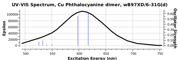
Andy Mclean posted a comment to my story of copper phthalocyanine (Monastral blue). The issue was its colour, and more specifically why this pigment has two peaks λmax 610 and 710nm making it blue. The first was accurately reproduced by calculation on the monomer, but the second was absent with such a model. Andy suggested this latter was due to stacking. Here, the calculated spectrum of a stacked dimer is explored.

Copper phthalocyanine, showing herring-bone stacking. Click for 3D.
The X-ray structure (above) shows layers of the phthalocyanine, dislocated so that the Cu of one unit aligns perfectly with a N of the units above and below the first one (Cu-N 3.28Å). This corresponds to the di-axially solvated system I
explored in a comment appended to the original post. The
TD-DFT calculated (since each unit is a doublet radical, the dimer was treated as a triplet state, this being much lower in energy than a singlet closed shell state) electronic spectrum for two units, stacked above each other as shown above reveals two transitions at ~ 600 and 620 nm. This is still some way away from reproducing the measured (solid state or solution spectra).
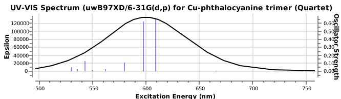
 The hypothesis must now be that the effect of such π-π stacking on the electronic spectrum converges only slowly with the degree of stacking (if indeed it is that that is the root cause of the 710nm transition). A calculation on a triple-layered model is currently under way (this being the absolute limit of what can be done without a periodic boundary model). The spectrum will be appended to this post in a week or so (see below). There is little sign of the spectrum evolving a quite separate band at 710nm. The model is still incomplete!
Tags:Andy Mclean, copper phthalocyanine, energy, Historical, TD-DFT, X-ray
Posted in Interesting chemistry | No Comments »
Monday, April 4th, 2011
Most scientific theories emerge slowly, over decades, but others emerge fully formed virtually overnight as it were (think Einstein in 1905). A third category is the supernova type, burning brightly for a short while, but then vanishing (almost) without trace shortly thereafter. The structure of DNA (of which I have blogged elsewhere) belongs to the second class, whilst one the brightest (and now entirely forgotten) examples of the supernova type concerns the structure of proteins. In 1936, it must have seemed a sure bet that the first person to come up with a successful theory of the origins of the (non-random) relatively rigid structure of proteins would inevitably win a Nobel prize (and of course this did happen for that other biologically important system, DNA, some 17 years later). Compelling structures for larger molecules providing reliable atom-atom distances based on crystallography were still in the future in 1936, and so structural theories contained a fair element of speculation and hopefully inspired guesswork (much as cosmological theories appear to have nowadays!).
Dorothy Wrinch was a mathematician who came up with just such a hypothesis for rigid protein structure, based in effect on elegance and symmetry, coupled with some knowledge of chemistry and crystallography[1]. She had noticed that the repeating polypeptide motif might be folded such that a cyclisation could occur to give what she termed a cyclol (an organic chemist would call this an aminol, and we would also now recognize it as a three-fold tetrahedral intermediate of the type involved in the hydrolysis of peptides). Wrinch proposed that this cyclisation could be repeated on a large scale to produce rigid scaffolds for proteins. The three-fold symmetric elegance of such motifs clearly appealed to this mathematician (the interesting symmetrical and conformational properties of the central cyclohexane-like ring were still to be fully recognised by anyone. Since Wrinch built many 3D models of her cyclols, one can but wonder how that central ring was represented, and whether its chair conformation was at all recognised. Another Nobel prize awaited the discoverer of this, Derek Barton).

The Cyclol structure. Click for 3D.
An immense controversy immediately broke out (not least because little direct spectroscopic evidence for the OH groups could be found). The story is rivetingly told by Patrick Coffey in his book Cathedrals of Science (ISBN 978-0-19-532134-0). Linus Pauling entered the fray in 1939[2], and one of the arguments he deployed was not so much symmetric elegance but thermodynamics (he also suggested hydrogen bonding and S-S linkages for rigidifying proteins). The proposed cyclisation, he suggested, led to a very high energy species. Whilst Wrinch attempted to refute this[3], Pauling’s arguments won almost everyone over. Although Wrinch forlornly continued to promote her idea, last reviewing the topic as late as in 1963[4], crystallography was now producing cast iron data for protein structures. None have ever emerged with a cyclol motif, and this hypothesis is now firmly consigned to untaught history[5]. To this day, no examples of the tris(aminol) cyclol ring are to be found in the Cambridge small molecule crystal structure database either (although some related tetrahedral intermediates are known as crystalline species, see for example here, and they can be quite easily characterised in solution, see for example[6].
When I read the story, it struck me that modern theory could easily verify how valid Pauling’s thermodynamic argument was. I have picked (ala)6 as my model, and have calculated the relative free energy (ΔG298) of the following three isomers.
- An acyclic zwitterionic form of this hexapeptide, calculated with a SCRF reaction field for water to allow for the ionic nature (ωB97XD/6-31G(d,p)), reveals a proton transfer to a neutral system, with an energy of +7.3 kcal/mol

Acyclic (ala)6, in zwitterionic form
- A cyclic neutral peptide, which results from elimination of water from 1, again calculated with a water reaction field (DOI: 10042/to-8219), revealing a relative free energy of +0.0 kcal/mol

Cyclic (ala)6
- The cyclic isomer 3 resulting from further cyclisation of 2 (DOI: 10042/to-8222) with a relative free energy of +69.0 kcal/mol

Cyclol model for (ala)6.
From this, it appears that model 3 is ~69 kcal/mol less stable than the cyclic peptide 2, or 11.6 kcal/mol per amino acid residue. Pauling’s thermodynamic arguments suggested a value of ~28 kcal/mol (a value which Wrinch disputed as unreliable). So, in one sense, the above calculation is closer to Wrinch than to Pauling! In another, it still means Wrinch was wrong!! It is worth speculating why Pauling’s estimate is out. The cyclol 3 exhibits anomeric stabilizations, which of course were unknown in Pauling’s time. Both 2 and 3 exhibit attractive, but different, van der Waals attractions which contribute to their stabilities. And Pauling took no account of any entropy differences between 2 and 3. In retrospect, 3 was simply too rigid to allow most enzyme catalysis models to function, as we recognise them nowadays.
You might ask why I have revived a long forgotten theory as the topic of this post. Well, I think it is always worth revisiting the past, and re-examining old assumptions. When we do so, we find that Wrinch did not miss by as much as her detractors perhaps implied. With a little more luck, she might have gotten it right. Science is a bit like that, you need a dose of luck sometimes!
References
- D.M. Wrinch, "The cyclol hypothesis and the “globular” proteins", Proceedings of the Royal Society of London. Series A - Mathematical and Physical Sciences, vol. 161, pp. 505-524, 1937. https://doi.org/10.1098/rspa.1937.0159
- L. Pauling, and C. Niemann, "The Structure of Proteins", Journal of the American Chemical Society, vol. 61, pp. 1860-1867, 1939. https://doi.org/10.1021/ja01876a065
- D.M. Wrinch, "The Geometrical Attack on Protein Structure", Journal of the American Chemical Society, vol. 63, pp. 330-333, 1941. https://doi.org/10.1021/ja01847a004
- D. WRINCH, "Recent Advances in Cyclol Chemistry", Nature, vol. 199, pp. 564-566, 1963. https://doi.org/10.1038/199564a0
- C. Tanford, "How protein chemists learned about the hydrophobic factor", Protein Science, vol. 6, pp. 1358-1366, 1997. https://doi.org/10.1002/pro.5560060627
- H.S. Rzepa, A.M. Lobo, M.M. Marques, and S. Prabhakar, "Characterizing a tetrahedral intermediate in an acyl transfer reaction: An undergraduate 1H NMR demonstration", Journal of Chemical Education, vol. 64, pp. 725, 1987. https://doi.org/10.1021/ed064p725
Tags:Cambridge, chair, Derek Barton, Dorothy Wrinch, energy, high energy species, Historical, mathematician, organic chemist, Patrick Coffey, relative free energy, thermodynamics, Tutorial material
Posted in Interesting chemistry | 2 Comments »
Saturday, March 5th, 2011
In this earlier post, I noted some aspects of the calculated structures of both Z- and B-DNA duplexes. These calculations involved optimising the positions of around 250-254 atoms, for d(CGCG)2 and d(ATAT)2, an undertaking which has taken about two months of computer time! The geometries are finally optimised to the point where 2nd derivatives can be calculated, and which reveal up to 756 all-positive force constants and 6 translations and rotations which are close to zero! This now lets one compute the thermodynamic relative energies using ωB97XD/6-31G(d) (for 2nd derivatives) and 6-31G(d,p) (for dispersion terms). All geometries are optimized using a continuum solvent field (water), and are calculated, without a counterion, as hexa-anions.
| Relative thermodynamic energies (kcal mol-1) of DNA duplexes. |
| system |
Total energy (duplex) |
Dispersion term |
ΔΔH298 |
Δ(-T.ΔS298) |
ΔΔG298 duplex |
ΔG298 single chain |
ΔΔG298 (Duplex) |
| Z-CGCG |
0.0 |
0.0 |
0.0 |
0.0 |
0.0 |
0.0 |
-60.3 |
| B-CGCG |
6.2 |
-4.2 |
8.0 |
3.9 |
11.9 |
+3.1 |
-54.7 |
| Z-ATAT |
0.0 |
0.0 |
0.0 |
0. |
0.0 |
0.0 |
-44.9 |
| B-ATAT |
-7.6 |
-12.8 |
-7.0 |
2.7 |
-4.3 |
-1.8 |
-45.7 |
Note how the CGCG duplex is more stable as a Z-helix, whilst the ATAT duplex prefers the B-helix. I will discuss the precise reasons for this elsewhere.
Tags:6-31G(d), ATAT duplex, B-DNA, CGCG duplex, dispersion energy corrections, energy, thermodynamic stability, Tutorial material, watoc11, wB97XD, Z-DNA
Posted in Interesting chemistry | 4 Comments »
Friday, January 21st, 2011
Much of chemistry is about bonds, but sometimes it can also be about anti-bonds. It is also true that the simplest of molecules can have quite subtle properties. Thus most undergraduate courses in chemistry deal with how to describe the bonding in the diatomics of the first row of the periodic table. Often, only the series C2 to F2 is covered, so as to take into account the paramagnetism of dioxygen, and the triple bonded nature of dinitrogen (but never mentioning the strongest bond in the universe!). Rarely is diberyllium mentioned, and yet by its strangeness, it can also teach us a lot of chemistry.
The diagram below is what many textbooks show. The diagram can vary (and hence confuse) slightly, in regard to the relative ordering of the σ and π energy levels originating from the overlap of the 2p orbitals. It depends on the atom, and for Be, the σ comes out higher than the π. The other key ordering is that the σ* antibonding orbital resulting from out of phase overlap of the two 2s orbitals is actually lower in energy than the π bonding orbital resulting from in-phase overlap of the 2p orbitals. Yes, an antibonding orbital is more stable than a bonding orbital!

Molecular orbital diagram for Be2
Well, the diagram shows that the pair of occupied molecular orbitals resulting from the two (symmetric and antisymmetric, or g and u) combinations of the 1s orbitals cancel each other, as do the 2s combinations, and we conclude the bond order for this molecule is zero! Actually, if a quantum mechanical calculation is performed (at the ωB97XD/6-311G(d,p) level), the bond length emerges as 2.81Å and a vibrational wavenumber of 167 cm-1 is predicted. Despite the zero bond order, a weak bond IS predicted, and this is the van der Waals or dispersion bond.
Let us now pump this molecule up to a higher energy state by a double excitation of the two electrons in the 2s σ* electrons. We have to split them up, one each, into the next available orbital, which is the π, to form a triplet state (just like di-oxygen).

The doubly excited state of diberyllium
Well, this (higher energy) state is certainly shorter (a contrast with my item on longer being stronger). The length is now 1.78Å, which is more than 1Å shorter than the original state, despite being ~ 45 kcal/mol higher in energy. The Be-Be stretching wavenumber goes up to 917 cm-1. With four electrons in bonding orbitals, diberyllium has a double bond! One can also pair the π electrons up to form an open shell (excited) singlet, which is ~ 51 kcal/mol higher than the closed shell (unbonded) singlet. This also has a length of 1.78Å and a marginally lower stretch of 909 cm-1. If you want to read more about the doubly excited state of this molecule, see DOI: 10.1139/v96-111.
One might be tempted to make an analogy between physics, and its particles and antiparticles. Yes, electrons can occupy antibonding as well as bonding orbitals. But the overall bond order will be reduced to zero if the total numbers of each are equal. And one can be pretty certain that there is no molecule at all in which the number of antibonding electrons exceeds the bonding ones! Or, if anyone is aware of such an example, do tell!
Tags:diberyllium, energy, excited state, higher energy, higher energy state, pence, Tutorial material
Posted in Interesting chemistry | 6 Comments »
Tuesday, December 7th, 2010
More inspiration from tutorials. In a lecture on organic aromaticity, the 4n+2/4n Hückel rule was introduced (in fact, neither rule appears to have actually been coined in this form by Hückel himself!). The simplest examples are respectively the cyclopropenyl cation and anion. The former has 2 π-electrons exhibiting cyclic delocalisation, and the 4n+2 (n=0) rule predicts aromaticity. Accordingly, all three C-C distances are the same (1.363Å).

Cyclopropenium cation and anion
The anion however appears to have 4 π-electrons, and must therefore belong to the 4n (n=1) rule and exhibit antiaromaticity. Pretty straight forward thus far. But students have a knack of asking apparently simple, but quite thought provoking questions. This one was “does one count lone pairs of electrons“? Perhaps a different way of putting it would be “does the lone pair really count as π-electrons?”
So, time for a calculation. Well, it turns out there are two isomers of the anion. The first has two C-C bond lengths of 1.383Å and one of 1.841Å; two short and one (very) long. Moreover, the whole system is very much non planar.

Cyclopropenium anion, first isomer
This isomer turns out to be really a 4π-allyl anion in disguise. To avoid any danger of cyclic conjugation (and hence antiaromaticity), the groups at the end of the allyl fragment rotate. So yes, this IS a 4π-electron system, but the molecule has cleverly distorted to avoid antiaromaticity as best it can.

Cyclopropenium anion. Isomer 2.
What about the second isomer? This now has one short (1.293Å) and two long (1.598Å) C-C lengths. The carbon bearing the two long bonds is now highly non planar. It is best described as an isolated double bond (2 π-electrons) trying to get as far away as possible, and to avoid as much overlap as it can, with a lone pair (NOT π) on the third carbon. Now, the lone pair really does NOT count, since it is too far from the other 2 π-electrons, and inclined at the wrong angle, to overlap effectively with them. The two isomers are almost the same in energy (the first being the lower in free energy by ~1 kcal/mol).
So what kind of answer would one give to the inquisitive tutee? Firstly, as the name implies, antiaromaticity is not good for a molecule. If it possibly can, it will avoid it. For the cyclopropenium anion, there are two quite effective ways of avoiding antiaromaticity. It is not, as a result, actually a good example of an antiaromatic system. Because molecules can be very clever at avoiding antiaromaticity, remarkably few examples of genuine antiaromatics actually exist!
I end with another way of looking at this problem using group theory. The cyclopropenium cation has D3h symmetry, and the LUMO (lowest unoccupied) molecule orbital in fact belongs to the E” irreducible representation. This means it is doubly degenerate. To form the anion from it, two electrons must be placed in one of these orbitals (but unless an open shell is formed, one cannot place one electron in each). Whichever orbital receives the two electrons is now stabilised, the degeneracy must break, and the resulting geometry must reflect this. The two symmetry-broken geometries are precisely those shown above.
 Cyclopropenium cation, E" LUMO orbital 11. Click for 3D |
 Cyclopropenium cation, E" LUMO orbital 12. Click for 3D |
Tags:energy, free energy, Huckel, Interesting chemistry, Pretty straight forward, Tutorial material
Posted in Interesting chemistry | 12 Comments »
Sunday, September 19th, 2010
Many university chemistry departments, and mine is no exception, like to invite applicants to our courses to show them around. Part of the activities on the day is an “interview” in which the candidate is given a chance to shine. Over the years, I have evolved questions about chemistry which can form the basis of discussion, and I thought I would share one such question here. It starts by my drawing on the blackboard (yes, I really still use one!) the following molecular structure.

Mystery molecule.
The candidate is then invited to offer their initial impressions of this molecule, and shortly thereafter asked how they might make it (or where perhaps they might be able to buy it). This of course floors even the most confident of applicants! But after a moments thought, most students can derive not only a molecular formula, but an empirical formula. From which it becomes apparent that it is actually a trimer of carbon dioxide. In the previous post, I showed the structure of solid CO2 and how an oxygen from one unit came fairly close to a carbon from another. So the next logical question might be to ask if this might lead to a molecule such as shown above. Why a trimer? Well, the aromatic core is also easily perceived, and one might expect some aromatic stabilization to result (which most of the candidates readily spot). Its also ionic, and here perhaps solvation may help stabilize. Finally, armed with Le Chatelier’s principle, one might conclude that pressure too would help. At this point of course, the realization normally dawns that possibly the purchase of a carbonated soft drink in a supermarket might offer perhaps a few molecules of the above. The discussion normally takes about 10 minutes, and is guaranteed to stimulate (and quite possibly exhaust) most students.
But here in this post, I would like to offer the denouement. What actually are the chances of forming this species? Enter a B3LYP/6-311G(d,p) calculation of the free energy. We can do this for various models:
- The trimer energy evaluated in a continuum solvent (water). This works out at 83.4 kcal/mol higher than three monomers (in part due to the entropic requirements of coalescing three molecules into one). So, not many molecules in a fizzy drink then! (just as well perhaps, since e.g. benzene as an aromatic molecule would not be a pleasant additive).
- How aromatic is the molecule? Well, a NICS(1) index of -2.3 ppm suggests little actual aromaticity. The C-O bond length (1.365Å) is certainly short enough. The Kekulé vibrational mode however is quite low (974 cm-1) compared to benzene, which is ~1310 cm-1 (remember, this mode represents how much energy it takes to distort an aromatic ring from a symmetric structure to the bond localized form).
- If its not aromatic, then perhaps after all a better representation might be:

A better resonance structure
It is worth asking why even this perfectly reasonable form is so much higher in free energy than carbon dioxide itself.
- Would solvating the structure with three explicit water molecules help (as per below)? Deciding quite how the hydrogen bonds will form is an interesting exercise in its own right!

Solvated trimer
But now the energy is +96.8 kcal/mol compared to the monomers. Its that entropy again!
- Actually, oxygen is pretty poor at propagating aromaticity. Nitrogen is much better, so what about the following (historically, such s-triazines were in fact much better known than benzene itself in the first half of the 19th century).

Carbo-diimide trimer
This is now merely +35.8 kcal/mol higher in free energy compared to three momers. The Kekulé mode is up to 1355 cm-1 (discuss!).
There are many other facets of this that could be raised. But the main reason for introducing such a molecule for discussion is that just by looking at the structure, so many ideas can be explored. That, by and large, is how chemistry works.
Tags:energy, free energy, Interesting chemistry, trimer energy
Posted in Interesting chemistry | 3 Comments »


